- Label
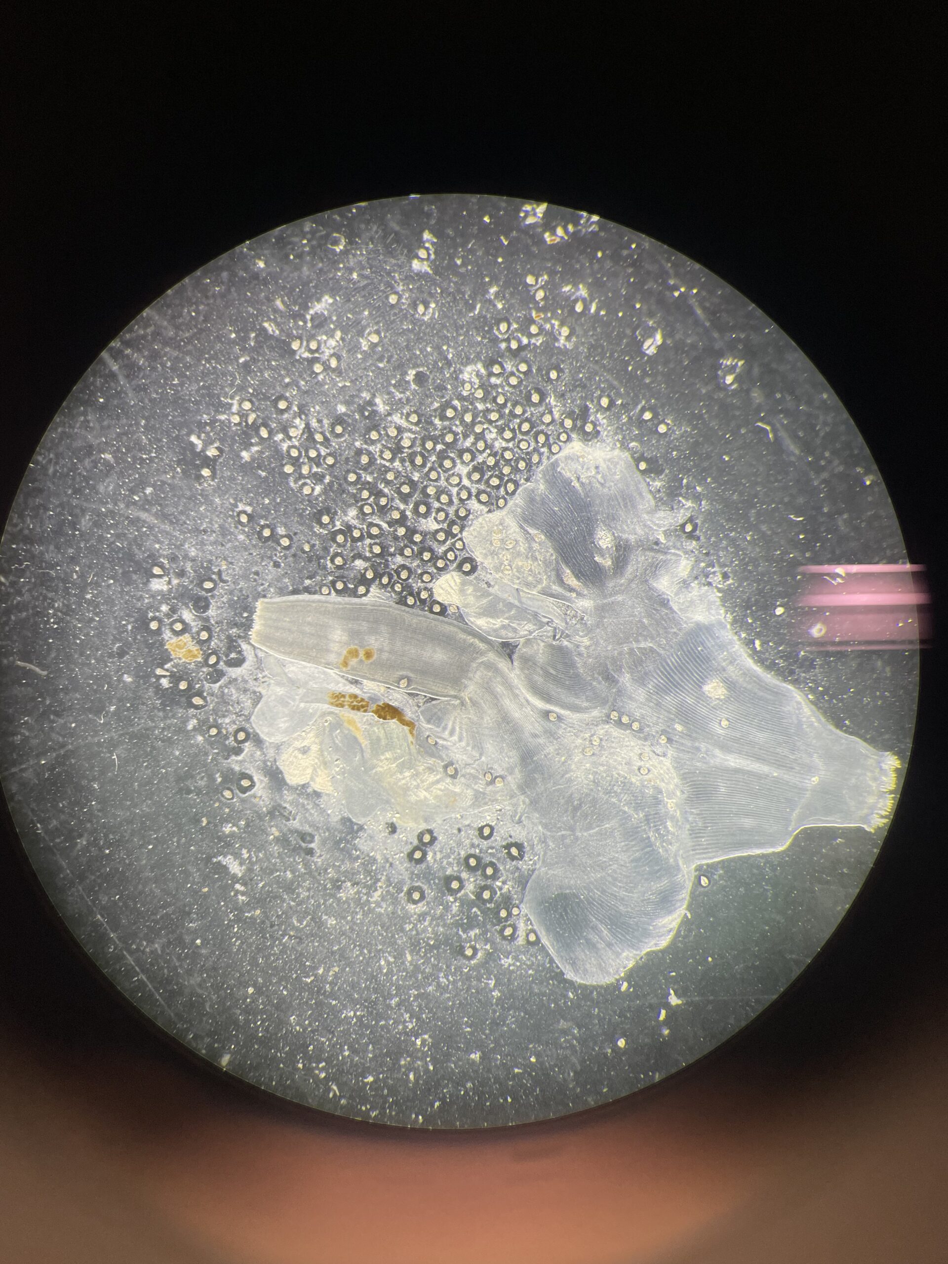
- Parasitized Clam
- Artist
- Sarah-Kate Srivastava
- Description
- Shown in the image is the siphon tissue of a coquina clam. It’s a first and second intermediate host to the trematode species Lasiotocus trachinoti before it moves to its definitive host, the Florida Pompano. The clam becomes infected by filtering out the parasite’s eggs from the water column. Sporocysts then form in the clam’s gonads, and many free swimming cercariae emerge and settle in the clam’s siphon tissue as metacercariae. When the clam is ingested by a fish, the metacercariae emerge as adults in the gut of the fish and the life cycle is complete. This particular clam’s tissue is heavily infected by the mesocercariae of this trematode species. Unlike most trematodes, this species is able to encyst and excyst in its host’s tissue, taking on two possible forms.
- Type
- Microscope
- Artist Affiliation
- Undergraduate Student
- Department
- Department of Biological Science
- Area of Research
- Aquatic Disease Ecology
- Research Group Site
- The Stewart Merrill RAD Lab
- Date
- 2024