Label
Odor in technicolor!
Artist
Auroni Khan
Description
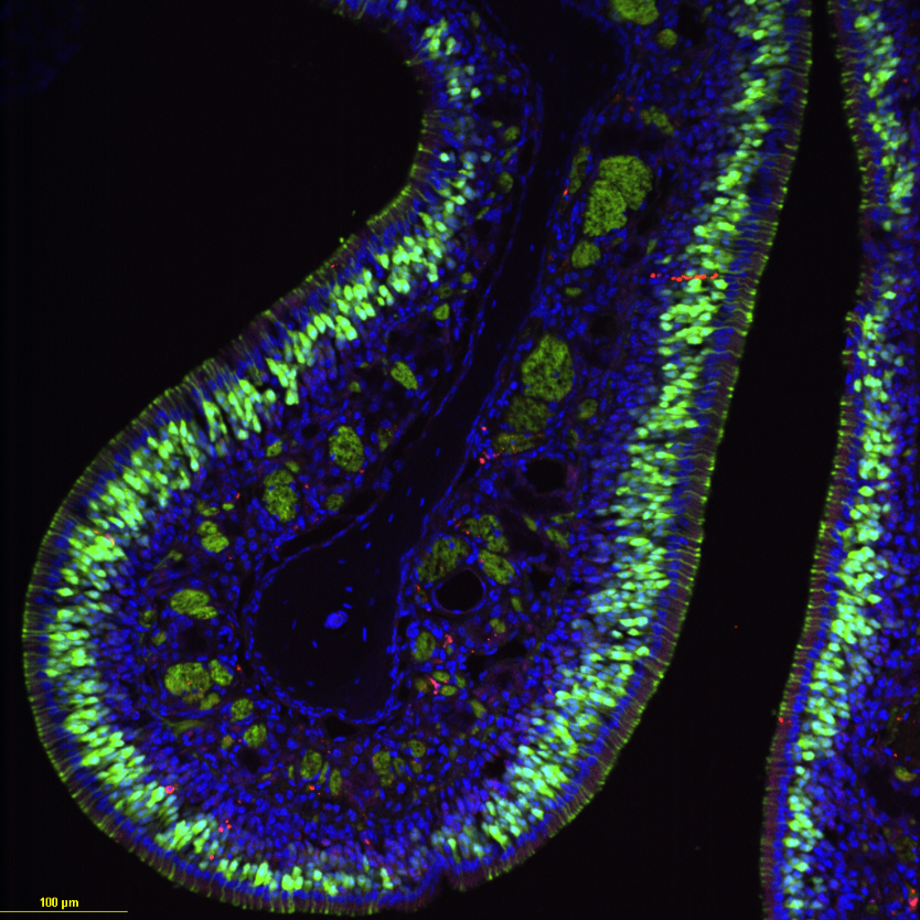
Mouse olfactory neurons after ablation with Methimazole and regeneration with Cannabidiol (CBD) treatment viewed using confocal microscopy at 20x. Green indicates neurons that are OMP-GFP (Olfactory Marker Protein-Green Fluorescent Protein) positive. Blue are all neurons viewed using DAPI, a dye that indicates nuclei in cells. Red are the neurons undergoing apoptosis indicated by Cas3.
Type
Microscope
Artist Affiliation
Graduate Student
Department
Department of Biological Science
Area of Research
Cell and Molecular Biology
Date
2024