Label
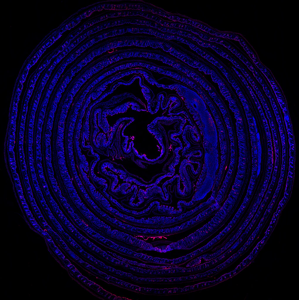
Unraveling the Second Brain
Artist
Abigail Randolph
Description
The large intestine of a Gcg-Cre/tdTomato reporter rat processed using an adapted Swiss Roll technique and counterstained with DAPI, a fluorescent dye. Native red fluorescence reveals the distribution and structure of gcg-expressing enteroendocrine cells, which are thought to be included in a population of neuropod cells that form synaptic connections with enteric neurons and vagal afferents. These cells play an essential role in transmitting information from the gut to the brain!
Type
Keyence BZ-X710 Microscope
Artist Affiliation
Graduate Student
Department
Department of Psychology
Area of Research
Neuroscience
Date
2023