Label
Multimodal Imaging of Harvester Ants
Artist
Elise Gonzalez
Description
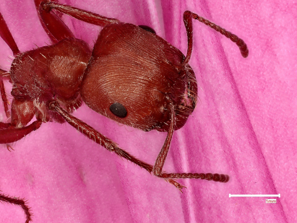
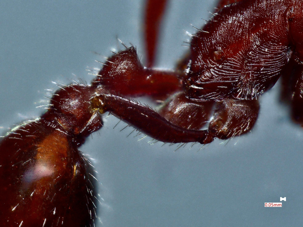
This art piece bridges the gap between science and art by utilizing a bio-aesthetic framework that treats the harvester ant as both a biological sample and a visual narrative. Unlike traditional photography that uses light, the scanning electron microscopy scanned the surface of the harvester ant and the flower petal with a focused beam of electrons. The left image was captured at 50x magnification and the right image at 80x magnification using a VHX digital microscope, which provides extreme depth of field and high-resolution structural detail to create a highly detailed, three-dimensional view of the specimens. This perspective shows the intricate, reddish-brown exoskeleton and compound eyes of the harvester ant — a species vital for seed dispersal in arid environments — set against a flower petal. The final composition was refined in Adobe Photoshop to enhance the visual storytelling of the biological sample.
Type
Microscope
Artist Affiliation
Undergraduate student
Department
Department of Biological Science
Area of Research
Microscopic biology art
Date
2026