- Label
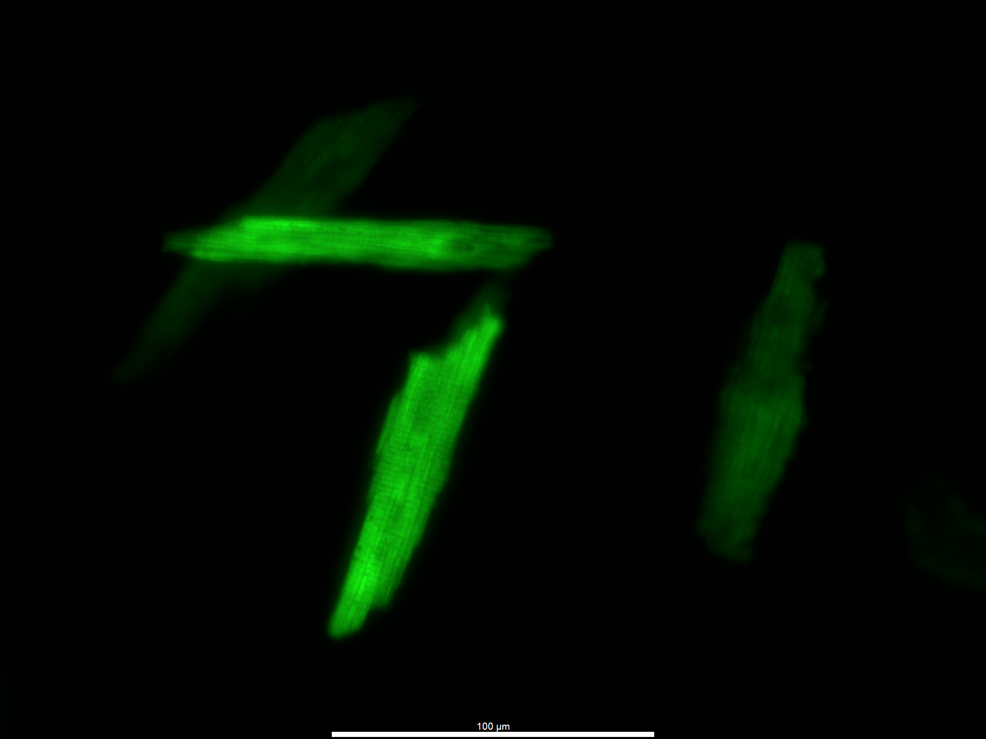
- Isolated Cardiomyocyte
- Artist
- Aida Rahimi Kahmini
- Description
- We are studying the effects of common substances that can impact human cardiovascular health. In this picture, we are showing a cardiomyocyte that has been exposed to nicotine, an active compound found in cigarette smoke. We used fluorescence imaging to capture the cardiomyocyte that was loaded with Fura-2 a calcium indicator dye. Sometimes I can see these single cells still beating under the microscope!
- Type
- Microscope
- Artist Affiliation
- Graduate Student
- Department
- Nutrition and Integrative Physiology
- Area of Research
- Cardiac Muscle and SSPN Protein
- Research Group Site
- Dr. Michelle Parvatiyar
- Date
- 2023