- Label
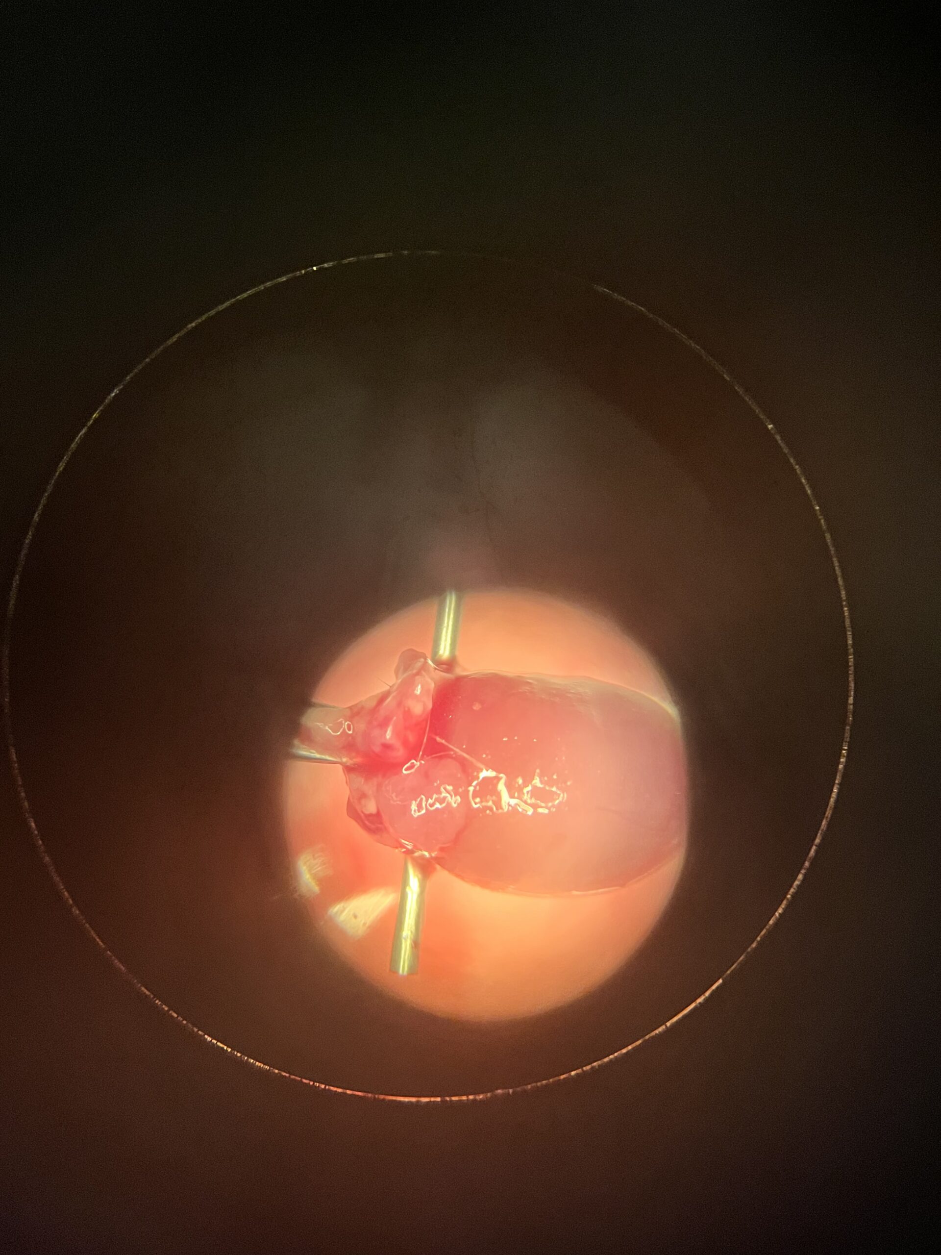
- Mouse Heart
- Artist
- Paula Nieto
- Description
- We study cardiomyopathies, diseases that affect the cardiac muscle, using mice models to study the pathogenesis of dilated cardiomyopathy and hypertrophic cardiomyopathy at the molecular, cellular and organ level. This picture was taken during a cardiomyocyte isolation experiment where we cannulate the aorta and perfuse the heart with an enzyme solution to ultimately obtain single cardiomyocyte cells. We can then measure calcium transients and contractility.
- Type
- Cell Phone Camera
- Artist Affiliation
- Graduate Student
- Department
- Biomedical Sciences
- Area of Research
- Cardiac Muscle
- Research Group Site
- Dr. Jose Pinto
- Date
- 2022