Label
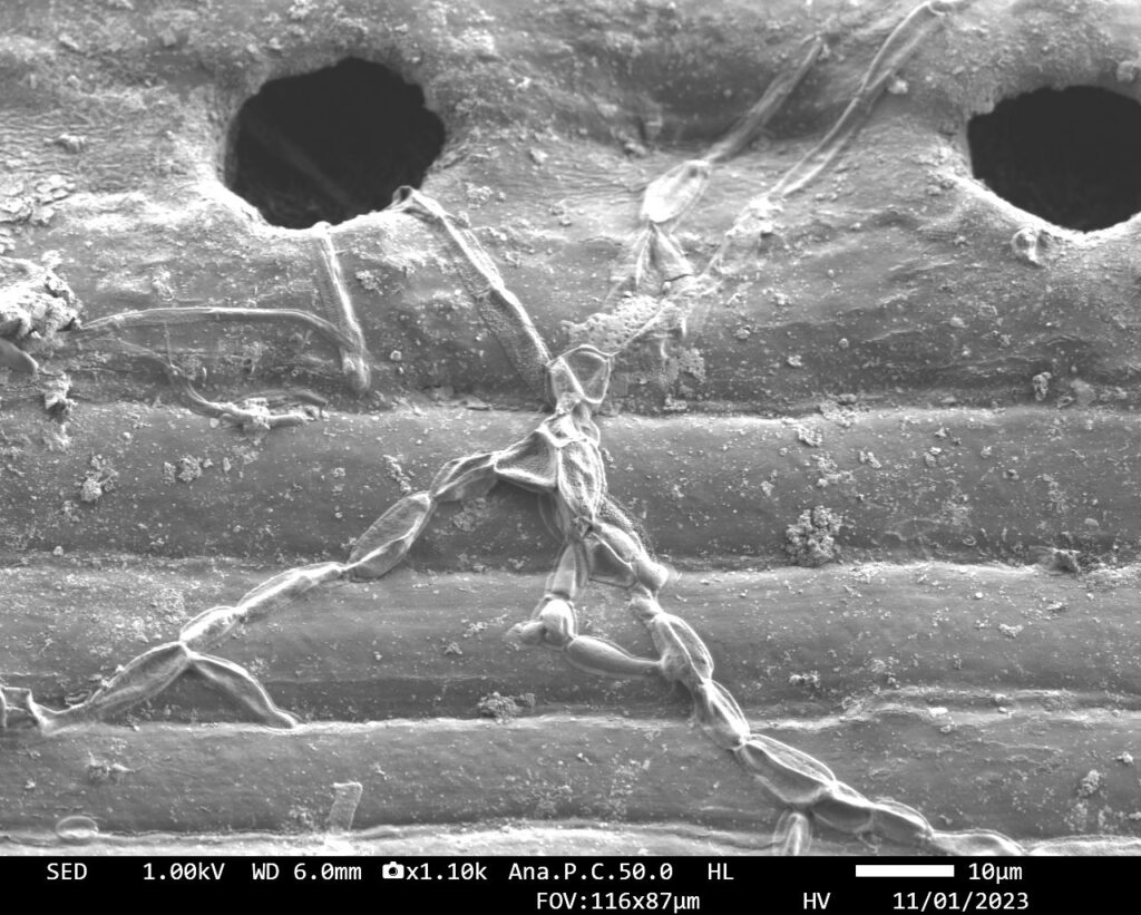
Microorganisms Investigating Stromata on Pine Needle
Artist
Catherine Fidd
Description
An image of the surface of a pine needle displaying a stroma (hole) on the surface of the needle. A tiny microorganism was captured sneaking one of its long tendrils into the stromata on the surface of the needle. A beautiful example of the synchronicity of life on the microscale.
Type
Scanning Electron Microscope
Artist Affiliation
Graduate Student
Department
Department of Materials Science and Engineering; Department of Biological Science
Area of Research
Tribology and Evolution
Date
2023