Tag: microscope
-
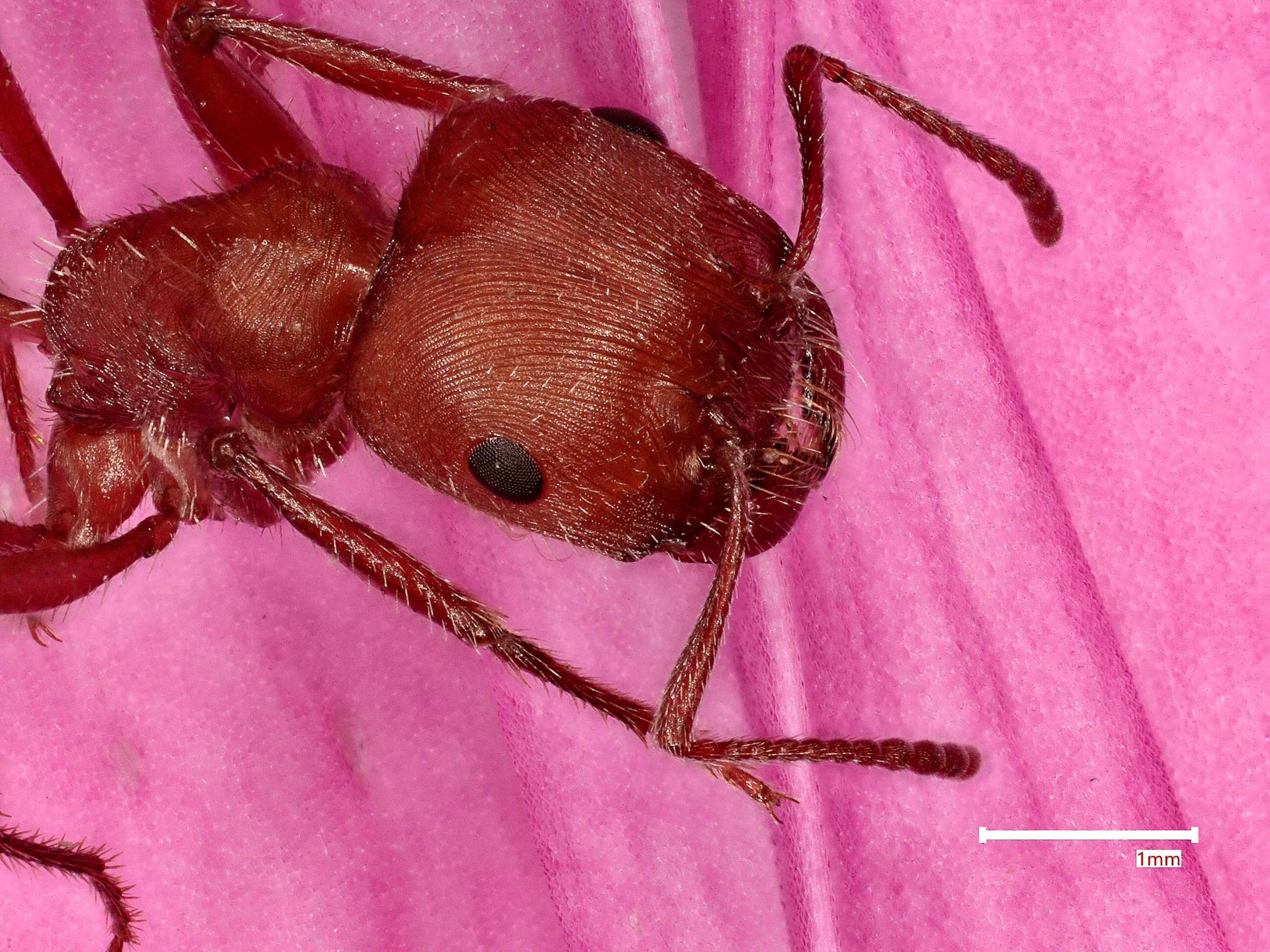
Multimodal Imaging of Harvester Ants
Label Multimodal Imaging of Harvester Ants Artist Elise Gonzalez Description This art piece bridges the gap between science and art by utilizing a bio-aesthetic framework that treats the harvester ant as both a biological sample and a visual narrative. Unlike traditional photography that uses light, the scanning electron microscopy scanned the surface of the harvester…
-
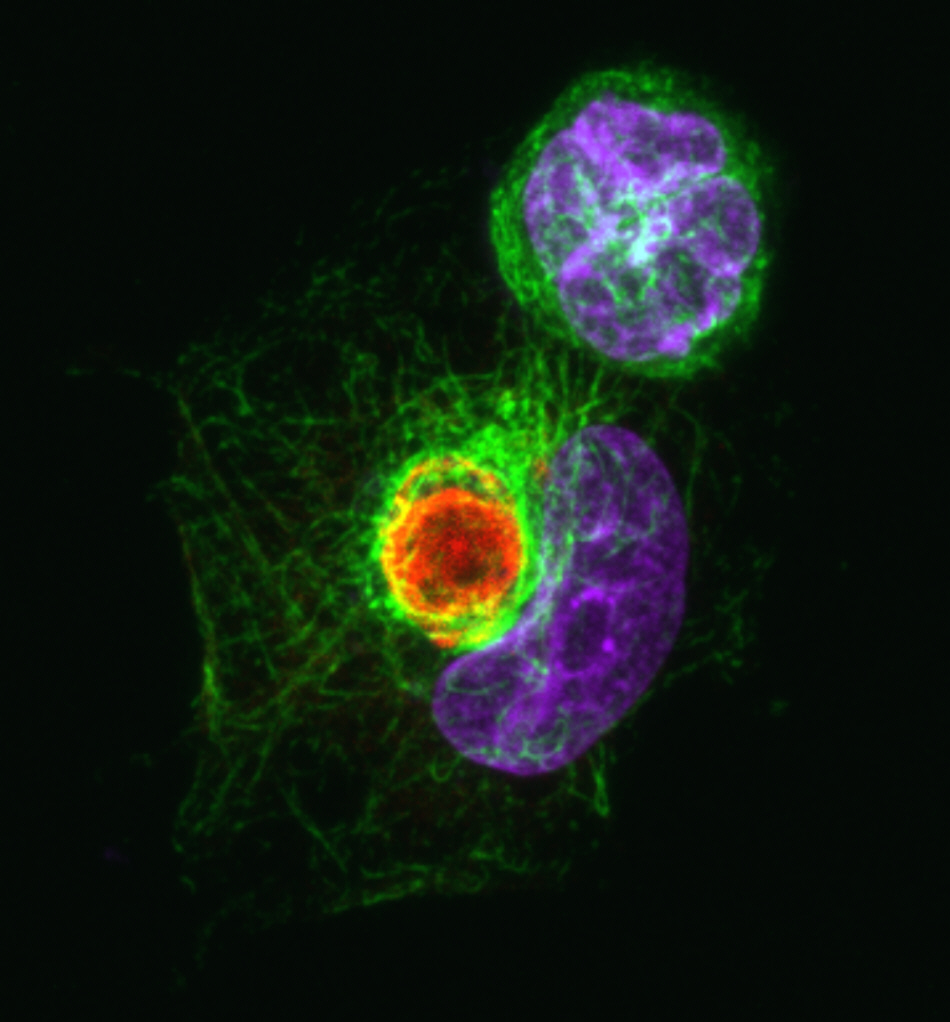
Zika Virus NS3 Drives the Assembly of the Viroplasm-Like Structure
Label Zika Virus NS3 Drives the Assembly of the Viroplasm-Like Structure Artist Tania Sultana Description Zika virus NS3 is a non-structural protein known to have protease and helicase activity. The image shows that NS3 is able to form a replication organelle or viroplasm-like structure (VLS) in human cells and microtubules rearrange around it. Red: NS3-driven…
-
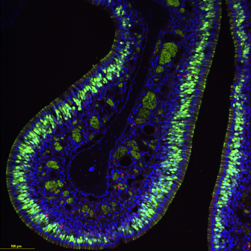
Odor in technicolor!
Label Odor in technicolor! Artist Auroni Khan Description Mouse olfactory neurons after ablation with Methimazole and regeneration with Cannabidiol (CBD) treatment viewed using confocal microscopy at 20x. Green indicates neurons that are OMP-GFP (Olfactory Marker Protein-Green Fluorescent Protein) positive. Blue are all neurons viewed using DAPI, a dye that indicates nuclei in cells. Red are…
-
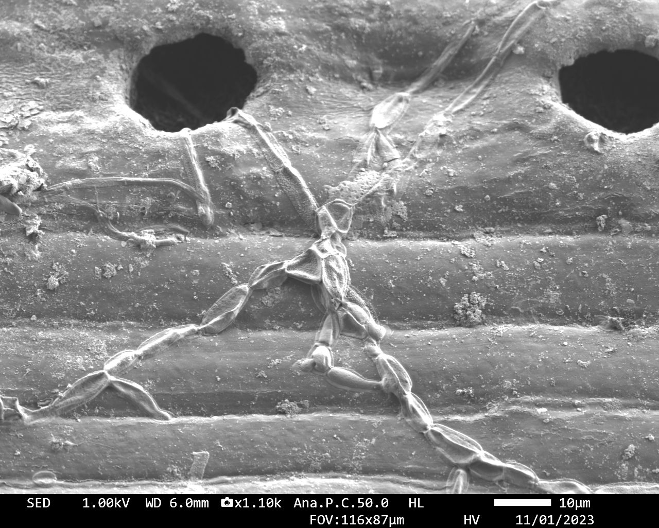
Microorganisms Investigating Stromata on Pine Needle
Label Microorganisms Investigating Stromata on Pine Needle Artist Catherine Fidd Description An image of the surface of a pine needle displaying a stroma (hole) on the surface of the needle. A tiny microorganism was captured sneaking one of its long tendrils into the stromata on the surface of the needle. A beautiful example of the…
-
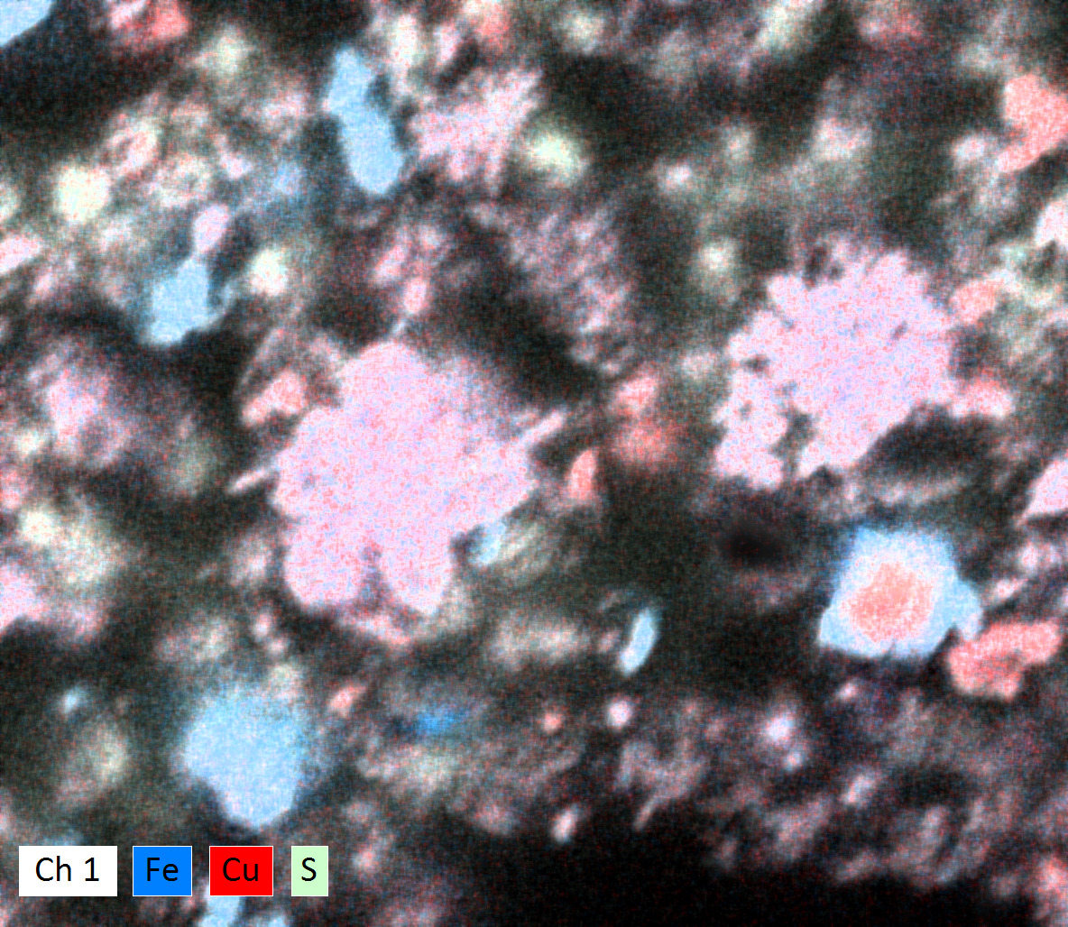
Echoes of Eden
Label Echoes of Eden Artist Binghui Li Description This is a cross-section scanning electron microscope (SEM) image of a micron-sized synthetic rock with the chemical formula CuFeS2 (chalcopyrite). Chalcopyrite is the most common copper-bearing mineral on Earth, and copper extraction is crucial due to increasing copper demand for electrification transition. Rock can be boring but…
-
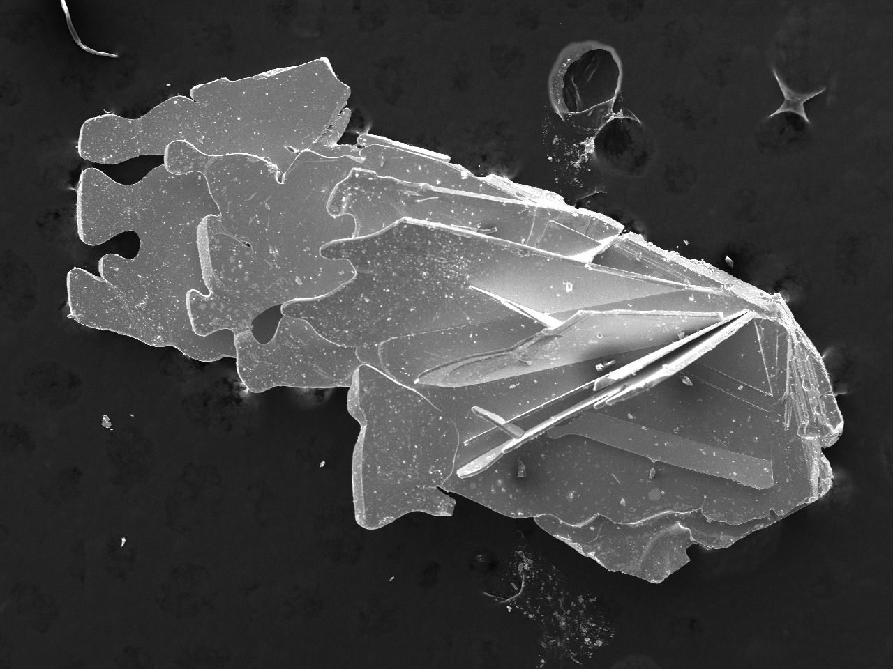
SEM Imaging of Polycrystalline Samples of Cr Intercalated TaTe2
Label SEM Imaging of Polycrystalline Samples of Cr Intercalated TaTe2 Artist David Saakov Description This image was created using the JOEL Scanning Electron Microscope in the Chemical Sciences Laboratory using 25kV accelerating voltage for higher resolution. The SEM is a high-powered microscope using electron beams to capture images of extremely small samples, in this case,…
-
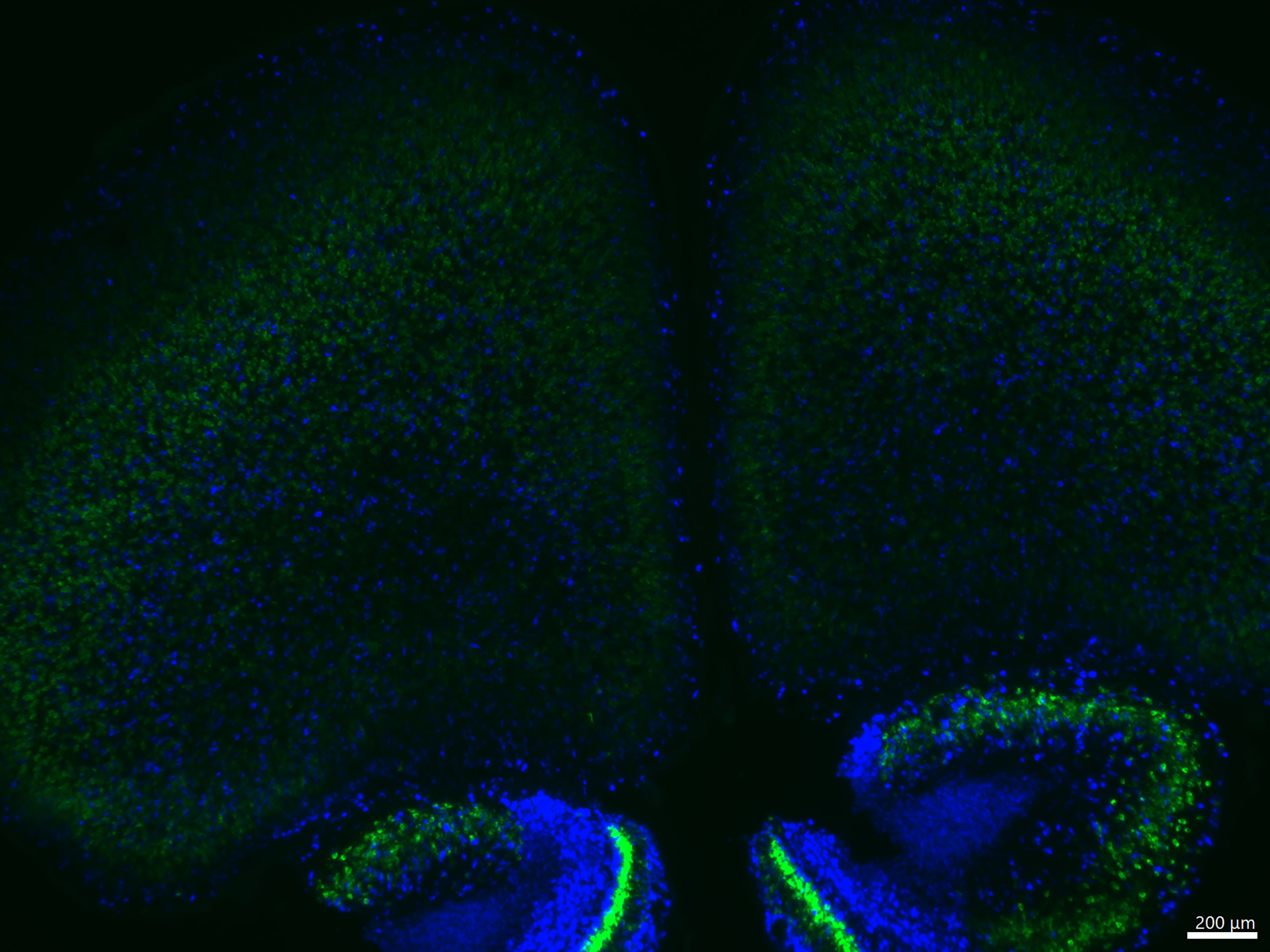
Excitatory and Inhibitory Neurons in the Developing Mouse Brain
Label Excitatory and Inhibitory Neurons in the Developing Mouse Brain Artist Yasmeena Thabet Description Fluorescent image showing a section of the mouse brain at postnatal day three, with excitatory neurons highlighted in green and inhibitory neurons marked in blue, illustrating the early organization of neuronal circuits. Type Microscope Artist Affiliation Graduate Student Department Department of…
-
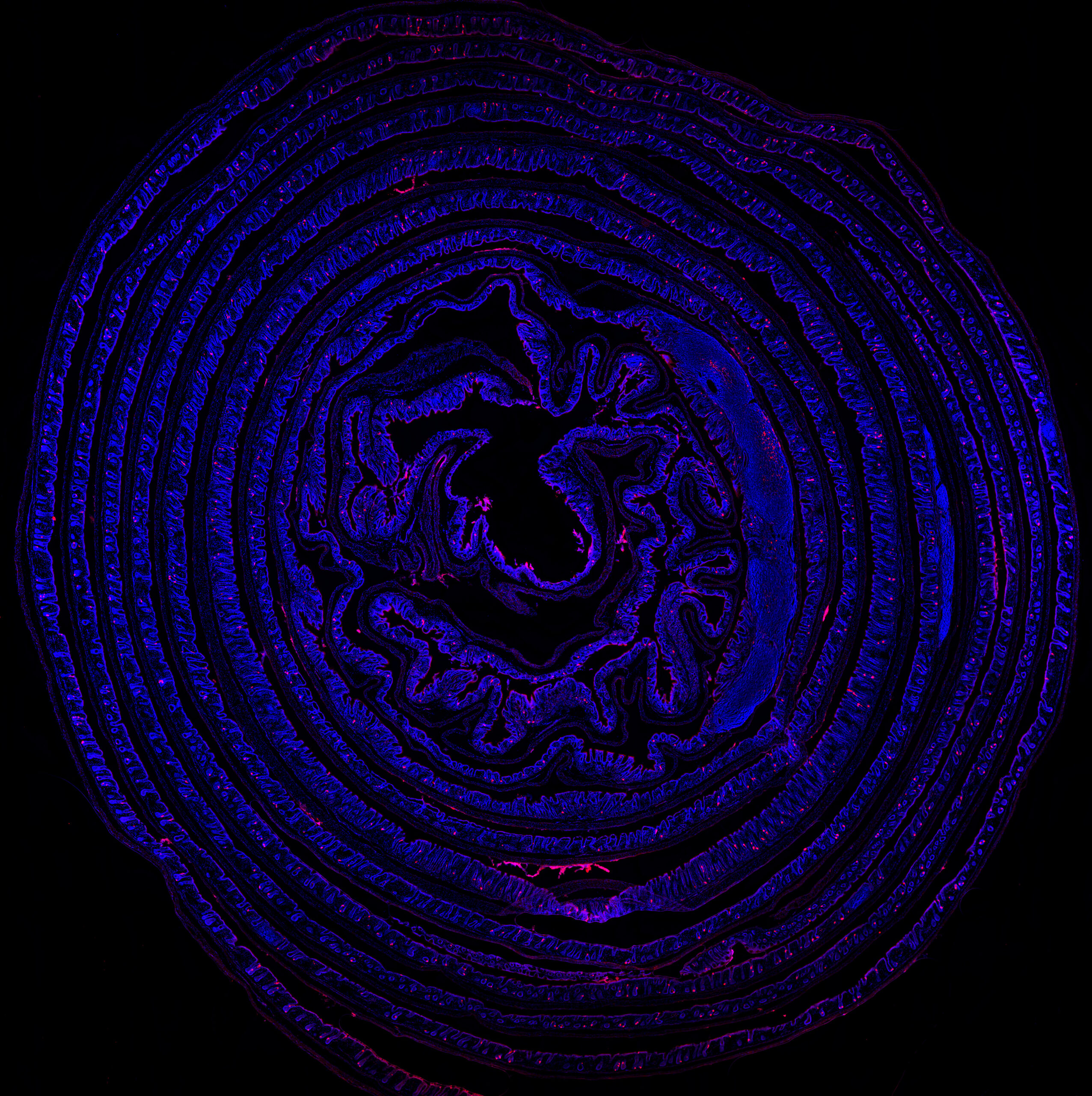
Unraveling the Second Brain
Label Unraveling the Second Brain Artist Abigail Randolph Description The large intestine of a Gcg-Cre/tdTomato reporter rat processed using an adapted Swiss Roll technique and counterstained with DAPI, a fluorescent dye. Native red fluorescence reveals the distribution and structure of gcg-expressing enteroendocrine cells, which are thought to be included in a population of neuropod cells…
-

Daphstronaut
Label Daphstronaut Artist David DuBose Description Just drifting through time. Type Microscope Artist Affiliation FSU Staff Department FSU Coastal and Marine Laboratory Area of Research Parasitology Research Group Site The Stewart Merrill RAD Lab Date 2023
-

Lifelike Illusions of Inorganic Microstructures
Label Lifelike Illusions of Inorganic Microstructures Artist Misa Amane Description This art features photographs of various silica-carbonate biomorphs, captured at 40x magnification with a Leica microscope and camera. Biomorphs are complex polycrystalline formations which arise from inorganic reactions. Research into these systems could lead to new crystallization methods and allow design of biomimetic materials with…